“My initial art was centered around trauma at the cellular level.”
Music vector created by macrovector – www.freepik.com
First, let me introduce myself. My name is Yana Zorina, and I am a cellular neuroscientist with a lifelong passion for the arts.
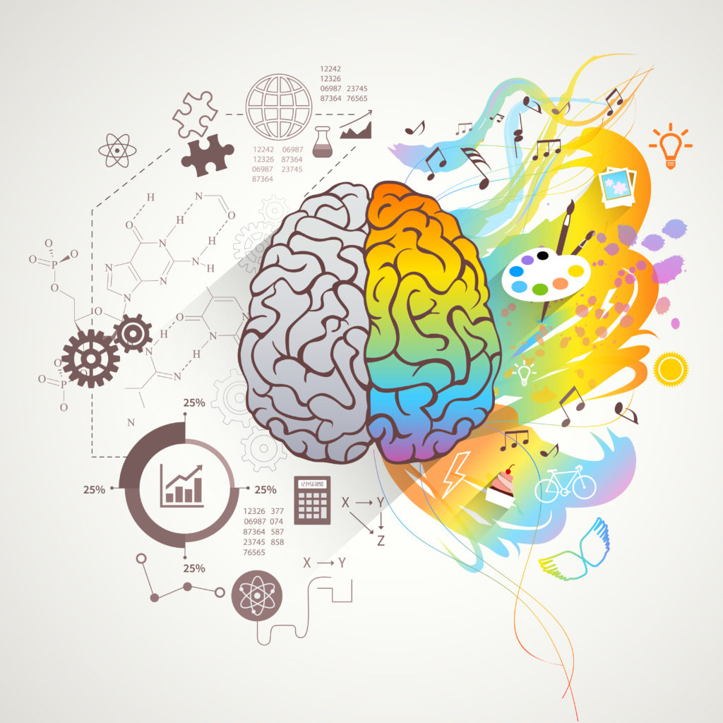
While the notion may be a bit outdated, it has traditionally been thought that logical thinking and scientific reasoning are primarily driven by the left hemisphere of the brain, whereas the right hemisphere has more creative and artistic inclinations. Even though this model is no longer considered to be anatomically correct, many of us can identify as being driven by one side more than the other.
Reflecting on my childhood, I spent most of my spare time immersing myself in intricate artwork and detailed craftsmanship, including anything from painting to crocheting and lacemaking. Paradoxically, in school I was primarily attracted to exact sciences such as mathematics, chemistry and later, not so exact biology. My brain strived to understand how the world works at the most basic level, invisible to the naked eye.
After toying around with the idea of becoming a professional artist, I decided to dedicate my career to my innate curiosity about how the human body works. As early as in middle school, I zoomed into understanding the mysterious labyrinth of the brain.
While the field of neuroscience has always attracted me due to the central role of the brain in controlling the rest of the body, I was also in for some pleasant surprises in studying the brain. It turned out that neurons (and glia) not only carry out complex functions, but they also have amazingly beautiful, complex morphology. With the development of state-of-the-art microscopy techniques, images of neurons and glia have taken center stage in adorning the walls of most Neuroscience departments in academic research centers across the country.
Confocal microscopy in particular has been near and dear to my heart throughout my graduate and postdoctoral training. The basic concept of confocal microscopy is focused on imaging a very thin optical slice of a biological sample. Despite focusing on a very thin section, neuronal images can reveal incredibly complex cellular morphology. When you combine this with multi-color fluorescence imaging, you also get an artistic rendering of these neuronal slices.
During my college and graduate school years, I somehow let my love of creating artwork slip to the back burner, but a few years ago I felt an acute need to bring it back. This became especially true after starting a family and having children. As a teenager, I spent a lot of time creating French beaded flowers. Later, while imaging immunofluorescently labeled cells [a process in which antibodies attached to dyes are used to bring color to specific types of cells or areas of cells], I began to think of the bright, colorful pixels in these images as beads. I thought that beads could be used to render the cellular structures observed under a microscope in 3 dimensions.
And this is where I found how my scientific and artistic sides could merge.
Art + Science
Art became a therapeutic practice for me, which I now use to let go of all the daily chaos and reach a state of “flow”. As initially defined by the Hungarian-American psychologist Mihaly Csikszentmihalyi, flow is “a state of concentration or complete absorption with the activity at hand and the situation. It is a state in which people are so involved in an activity that nothing else seems to matter.” Such absorption can come from work that is very intellectually stimulating, as well as a rhythmic and repetitive processes, such as knitting.
While my initial intention was just to show the beauty of fluorescence microscopy images in 3D renderings with beads, beadwork became my emotional oasis. Perhaps for this reason, I find myself today putting deeper meaning into the images I create. These works have begun to serve as metaphors for life experiences, such as stages of loss, grief and hope.
I often wonder how much it matters whether the message that the artist puts into their work is the same one that is perceived by the viewers. History tends to repeat itself, at both global and very personal levels, allowing us to put new meaning into art. Since my scientific research was focused on acute injury to the central nervous system, my initial art was centered around trauma at the cellular level. But several years later, this work came to represent a painful personal experience. And in the present day, the whole world is going through a major tragedy as the COVID-19 pandemic takes its toll.
Can art help us put our feelings in context and validate them?
The nervous system is very fragile and difficult to restore. Yet some people show extraordinary resilience to both physical and mental trauma. They retain their connection with hope, which is often so hard to find. In my artwork, I explore how at its most basic level, the nervous system can experience shock and adapt to changing circumstances.
We have built-in resilience, which allows us to move past obstacles, find hope in novel medical approaches and remain optimistic. These qualities are mirrored in nerve cells being able to move past an injury site, which I explored in one of my favorite works titled “Tortured”. This work is directly based on my postdoctoral research of neuronal regeneration and I would like to use it as an introduction to my SciArt practice.
Here is the story behind it.
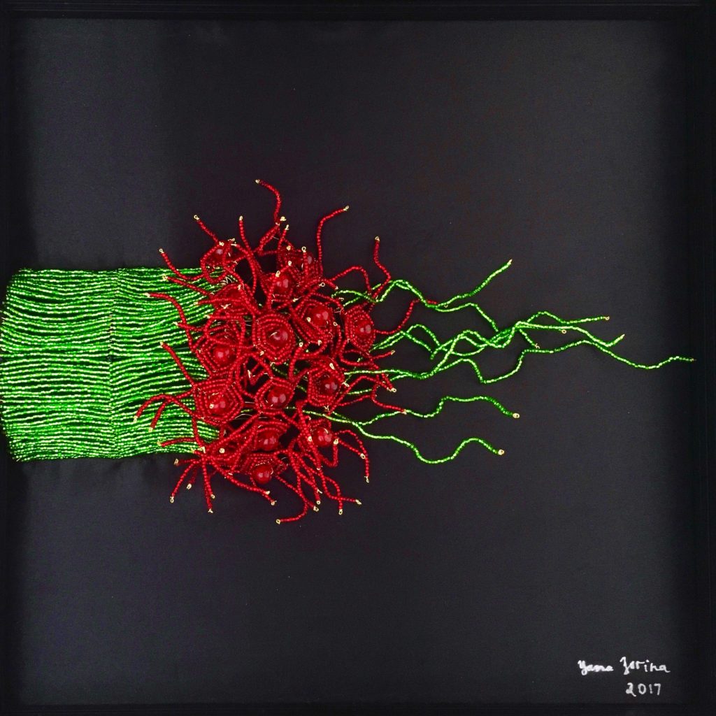
“Tortured”, 2017 – 3-dimensional beadwork representing nerve injury
Traumatic injury to the central nervous system results in an inhibitory environment. This condition presents a serious challenge for neurons that attempt to regenerate. In addition to inhibitory biochemical signals, a physical barrier called a “glial scar” forms at the injury site. Very few cells succeed in extending their axons beyond the glial scar. In contrast to healthy cells, axons that manage to pass the scar show a very distorted or “tortured” morphology. This phenomenon provides a metaphorical representation of a traumatic life event. People may be able to move forward, but they are no longer the same as before.
Please check out more of my artwork at www.neurobead.com.
Featured image: Sunrise – Where intricate beadwork meets precision of confocal microscopy.